CHRONIC EOSINOPHILIC LEUKEMIA (CEL)
Vysis 4q12 Tri-Color Rearrangement FISH Probe Kit
For more information, contact Abbott.
For more information, contact Abbott.


| PRODUCT NAME | UNIT SIZE | ORDER NUMBER | GTIN |
|---|
The Vysis 4q12 Tri-Color Rearrangement FISH Probe Kit is intended to detect rearrangements in chromosome 4q12 involving the FIP1L1- PDGFRA region, using the fluorescence in situ hybridization (FISH) technique.
The SpectrumGreen probe spans approximately 703 kb (chr4:53159272- 53862621; March 2006 assembly) and is located centromeric to the FIP1L1gene region. The SpectrumOrange probe spans approximately 448 kb (chr4:54045936-54494304; March 2006 assembly) and is located between the FIP1L1 and the CHIC2 gene regions. The SpectrumAqua probe spans approximately 578 kb (chr4:54840090- 55418505; March 2006 assembly) and extends from the telomeric end of the PDGFRA gene region to beyond the KIT gene region.
FISH signal patterns in nuclei having interstitial deletions of the orange probe target on one chromosome 4 homolog should be observed as one tri-color fusion and one green/aqua fusion lacking an orange signal. If the intervening orange probe target is not deleted, but relocated to another separate chromosomal location, the expected pattern would be one tri-color fusion, one green/aqua fusion and one lone orange signal. In instances of translocations involving the PDGFRA gene with loci on other chromosomes, the expected signal pattern would be one tri-color fusion, one orange/green fusion, and one separate aqua signal.
In interphase nuclei of normal cells, the probe is expected to appear as two tri- color (green, orange,aqua) fusions. In these fusions, overlapping orange and green signals may be perceived as yellow fusion signals with appropriate filters.
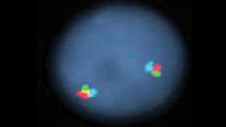
Normal Hybridization: Normal nucleus showing the two tricolor green/orange/aqua fusion signals.
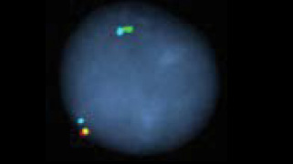
Abnormal Hybridization: Abnormal nucleus showing the one tricolor green/ orange/aqua fusion signal and one green/ aqua fusion signal with the orange signal deleted.

Please be aware that the website you have requested is intended for the residents of a particular country or countries, as noted on that site. As a result, the site may contain information on pharmaceuticals, medical devices and other products or uses of those products that are not approved in other countries or regions.
Please be aware that the website you have requested is intended for the residents of a particular country or countries, as noted on that site. As a result, the site may contain information on pharmaceuticals, medical devices and other products or uses of those products that are not approved in other countries or regions.