For more information, contact Abbott.
For more information, contact Abbott.
| PRODUCT NAME | UNIT SIZE | ORDER NUMBER | GTIN |
|---|
The Vysis BCR/ABL1/ASS1 Tri-Color DF FISH Probe Kit is intended to detect the t(9;22)(q34;q11.2) reciprocal translocation involving the BCR and ABL1 gene regions using the fluorescence in situ hybridization (FISH) technique.
The t(9;22) translocation which fuses the BCR gene on chromosome 22q11.2 and the ABL1 gene on chromosome 9q34 is observed by cytogenetics in greater than 80% of patients with chronic myelogenous leukemia (CML). In CML cases lacking a cytogenetically detectable translocation, the BCR/ABL1 fusion can still almost always be detected by FISH or other molecular techniques. BCR/ ABL1 fusions also occur in a portion of acute lymphocytic leukemia cases and more rarely in acute myeloid leukemia. In about 15 to 20 percent of CML cases, the t(9;22) results in the loss of genetic material flanking the BCR and/or ABL1 breakpoints on the derivative 9 chromosome. This loss can prevent the production of the highly specific two-fusion signal patterns expected of dual fusion probes and balanced translocations. If both BCR and ABL1 targets are deleted on the der(9) chromosome, low-level random overlap of orange and green signals within normal cells (producing a 1 orange, 1 green, 1 fusion pattern) cannot be discriminated from low-level true BCR/ABL1 fusions producing the same pattern. The Tri-Color design of this test uses a probe in a third color (aqua) on the centromeric side of the ABL1 breakpoint, which co-localizes with the orange signal in a random orange/green signal fusion, but is absent from a true BCR/ABL1 molecular fusion on the der(22) chromosome. The probes in this kit have been used in published papers to detect low levels of positive cells in CML patients who were undergoing therapy and had deletions of FISH signals on the derivative chromosome 9.
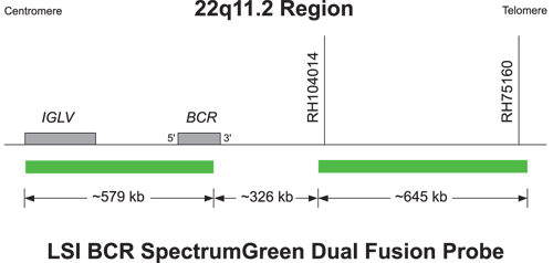
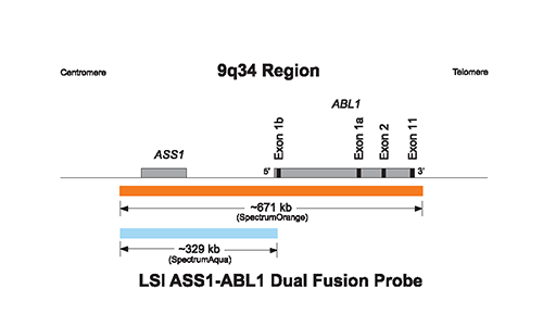
The approximately 671 kb (chr9:132255025-132926107; March 2006 assembly, UCSC Genome Browser) SpectrumOrange LSI ABL1 probe spans the ABL1 and ASS1 genes on chromosome 9q34. The approximately 329 kb (chr9:132255025-132584487; March 2006 assembly, UCSC Genome Browser) SpectrumAqua LSI ASS1 probe overlays with part of the area covered by the SpectrumOrange probe, spans the ASS1 gene and lies centromeric to the ABL1 gene breakpoint regions. The SpectrumGreen LSI BCR probe consists of two probes located at chromosome 22q11.2. The centromeric segment of the SpectrumGreen probe is approximately 579 kb (chr22:21382633-21962088 March 2006 assembly), and contains the majority of the BCR gene. The telomeric segment of the SpectrumGreen probe is approximately 645 kb (chr22:22288218-22932815; March 2006 assembly), and it lies telomeric to the BCR gene breakpoint region. There is an approximate 326 kb gap between the two green probes.

Normal Hybridization: Nucleus showing the two aqua/orange and two green signal pattern
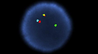
Abnormal Hybridization: Nucleus showing the one aqua/orange, one green, and one orange/green fusion (yellow) signal pattern.

Please be aware that the website you have requested is intended for the residents of a particular country or countries, as noted on that site. As a result, the site may contain information on pharmaceuticals, medical devices and other products or uses of those products that are not approved in other countries or regions.
Please be aware that the website you have requested is intended for the residents of a particular country or countries, as noted on that site. As a result, the site may contain information on pharmaceuticals, medical devices and other products or uses of those products that are not approved in other countries or regions.