SARCOMAS
Vysis DDIT3 Break Apart FISH Probe Kit
For more information, contact Abbott.
For more information, contact Abbott.
| PRODUCT NAME | UNIT SIZE | ORDER NUMBER | GTIN |
|---|
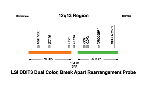
This fluorescence in situ Hybridization (FISH) probe is intended to detect chromosome rearrangements involving the DDIT3 (CHOP) gene located on chromosome 12q13.
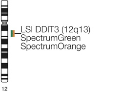
The Vysis LSI DDIT3 (12q13) Dual Color, Break Apart Rearrangement Probe consists of a mixture of 2 FISH DNA probes. The first probe, an ~700 kb probe labeled in SpectrumOrange lies proximal to the DDIT3 gene. The second probe labeled in SpectrumGreen extends distally from the DDIT3 gene and is ~663 kb in length.
In a normal cell that lacks a t(12q13) in the DDIT3 gene region, a two fusion signal pattern will be observed which reflects the two intact copies of DDIT3.
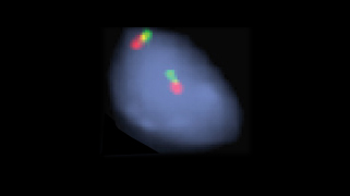
In an abnormal cell with a simple t(12q13), a one fusion, one green, and one orange signal pattern will be expected.
Normal Hybridization: Result of the hybridization of the Vysis LSI DDIT3 (12q13) Dual Color, Break Apart Rearrangement Probe, showing the two fusion signal pattern as observed in normal interphase cells.

Abnormal Hybridization: Abnormal cells hybridized with the Vysis LSI DDIT3 (12q13) Dual Color, Break Apart Rearrangement Probe. Two of the cells in this image show the one fusion, one orange, and one green signal pattern indicative of a rearrangement of one copy of the DDIT3 gene region.

Please be aware that the website you have requested is intended for the residents of a particular country or countries, as noted on that site. As a result, the site may contain information on pharmaceuticals, medical devices and other products or uses of those products that are not approved in other countries or regions.
Please be aware that the website you have requested is intended for the residents of a particular country or countries, as noted on that site. As a result, the site may contain information on pharmaceuticals, medical devices and other products or uses of those products that are not approved in other countries or regions.