For more information, contact Abbott.
For more information, contact Abbott.
| PRODUCT NAME | UNIT SIZE | ORDER NUMBER | GTIN |
|---|
The Vysis ETV6/RUNX1 DF FISH Probe Kit is intended to detect the t(12;21) (p13;q22) translocation between the ETV6 gene and the RUNX1 gene using the fluorescence in situ hybridization (FISH) technique.
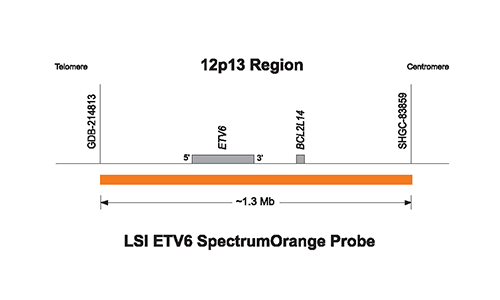
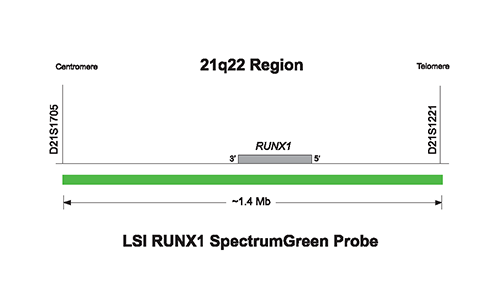
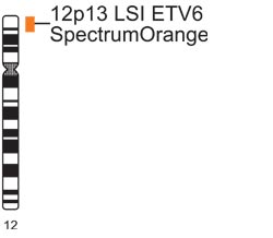
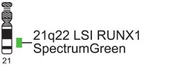
The approximately 1.3 Mb (chr12:11321286-12578034; March 2006 assembly) SpectrumOrange probe spans the ETV6 breakpoint region. The approximately 1.4 Mb (chr21:34452353-35813329; March 2006 assembly) SpectrumGreen probe spans the RUNX1 breakpoint region.
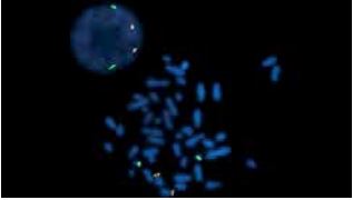
Normal Hybridization: The above image show two lymphocyte cells, one in interphase (upper left) and one in metaphase cell (lower right), that have been hybridized with the LSI ETV6/ RUNX1 Dual Color Dual Fusion Probe. Both cells show the two orange (RUNX1), two green (ETV6) signal pattern.
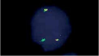
Abnormal Hybridization: The above image shows a bone marrow cell in interphase hybridized with the LSI ETV6/RUNX1 Dual Color Dual Fusion Probe. The cell in this image shows the one orange (RUNX1), one green (ETV6), two fusion (der (12) and der (21)) signal pattern.

Please be aware that the website you have requested is intended for the residents of a particular country or countries, as noted on that site. As a result, the site may contain information on pharmaceuticals, medical devices and other products or uses of those products that are not approved in other countries or regions.
Please be aware that the website you have requested is intended for the residents of a particular country or countries, as noted on that site. As a result, the site may contain information on pharmaceuticals, medical devices and other products or uses of those products that are not approved in other countries or regions.