SARCOMAS
Vysis FUS Break Apart FISH Probe Kit
For more information, contact Abbott.
For more information, contact Abbott.
| PRODUCT NAME | UNIT SIZE | ORDER NUMBER | GTIN |
|---|
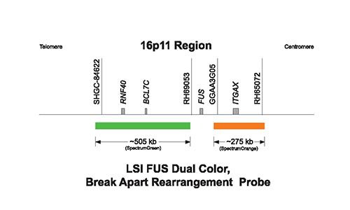
This fluorescence in situ hybridization (FISH) probe is intended to detect chromosome rearrangements involving the FUS gene located on chromosome 16p11.
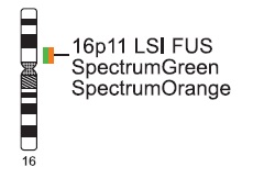
The Vysis LSI FUS (16p11) Dual Color, Break Apart Rearrangement Probe is a mixture of 2 probes. The first probe, a 505 kb probe labeled in SpectrumGreen, lies distal to the FUS gene. The second probe, labeled in SpectrumOrange, extends proximally from the FUS gene and is 275 kb in length.
The anticipated signal pattern in abnormal cells having a chromosomal breakpoint within the gap between the two probe targets on one chromosome 16 is one orange, one green, and one fusion signal. Other patterns may be observed if additional genetic alterations are present. Hybridization of this probe to interphase nuclei of normal cells is expected to produce two pair of overlapping, or nearly overlapping, orange and green (yellow fusion) signals.

Normal Hybridization: Normal cell hybridization using the Vysis LSI FUS (16p11) Dual Color, Break Apart Rearrangement Probe.
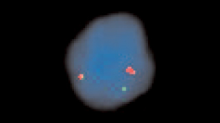
Abnormal Hybridization: Abnormal cell hybridization using the Vysis LSI FUS (16p11) Dual Color, Break Apart Rearrangement Probe.

Please be aware that the website you have requested is intended for the residents of a particular country or countries, as noted on that site. As a result, the site may contain information on pharmaceuticals, medical devices and other products or uses of those products that are not approved in other countries or regions.
Please be aware that the website you have requested is intended for the residents of a particular country or countries, as noted on that site. As a result, the site may contain information on pharmaceuticals, medical devices and other products or uses of those products that are not approved in other countries or regions.