Vysis LSI IGH/CCND1 DF FISH Probe Kit
For more information, contact Abbott.
For more information, contact Abbott.
| PRODUCT NAME | UNIT SIZE | ORDER NUMBER | GTIN |
|---|
These fluorescence in situ hybridization (FISH) probes are intended to detect the t(11;14)(q13;q32) reciprocal translocation involving the IGH and CCND1 gene regions.
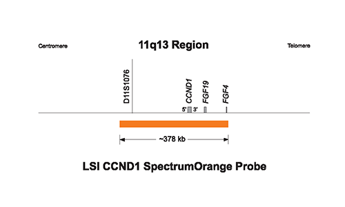
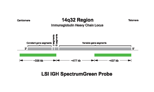
The approximately 378 kb SpectrumOrange probe spans the CCND1 gene (chr11:68927577-69305335; March 2006 assembly, UCSC Genome Browser). The SpectrumGreen probe consists of 2 probes; 1 IGH probe hybridizes to an approximately 338 kb region spanning the IGH constant region (chr14:105087705-105425263; March 2006 assembly, UCSC Genome Browser) and the other IGH probe is approximately 437 kb and hybridizes to the telomeric portion of the IGH variable region (chr14:105901997-106339460; March 2006 assembly, UCSC Genome Browser). There is an approximate 477 kb gap between the 2 probes.
LSI IGH/CCND1 hybridized to a cell containing t(11;14) with breakpoints at the MTC on 11q13 and at the IGH J region on 14q32 is expected to result in a signal pattern of two orange/green (yellow) fusions, one on each of the abnormal chromosomes 11 and 14 and single orange and green signals from the normal chromosomes.
Due to the gap between the two probes in the IGH probe set, the normal IGH loci may sometimes appear as two slightly separated green signals. This gap may also cause a slight separation of the orange and green signals on the der(11) chromosome, in some instances. Analysis of t(11;14) samples suggests that due to variation in breakpoint location on 11q13 loss of V segments within the LSI IGH probe target, some samples containing t(11;14) might display signal patterns different than 1O1G2F.
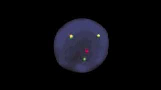
Abnormal Hybridization: LSI IGH/CCND1 Dual Color, Dual Fusion Translocation Probe hybridized to an abnormal nucleus showing the common 1O1G2F signal pattern.

Please be aware that the website you have requested is intended for the residents of a particular country or countries, as noted on that site. As a result, the site may contain information on pharmaceuticals, medical devices and other products or uses of those products that are not approved in other countries or regions.
Please be aware that the website you have requested is intended for the residents of a particular country or countries, as noted on that site. As a result, the site may contain information on pharmaceuticals, medical devices and other products or uses of those products that are not approved in other countries or regions.