Vysis LSI IGH/CCND1 XT Dual Color Dual Fusion FISH Probe Kit
For more information, contact Abbott.
For more information, contact Abbott.
_176kb_219kb_455kb_probemap_mw002-orange-copy-(1).png)

| PRODUCT NAME | UNIT SIZE | ORDER NUMBER | GTIN |
|---|
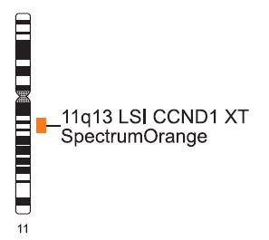
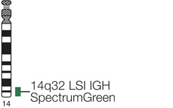
These fluorescence in situ hybridization (FISH) probes are intended to detect t(11;14)(q13;q32) reciprocal translocation involving the IGH and CCND1 gene regions.
The approximately 942 kb SpectrumOrange probe spans the CCND1 breakpoint region with gaps of 30 kb and 62 kb. The contig is composed of 3 segments of approximately 176 kb (chr11:68363475-68539031; UCSC March 2006), 219 kb (chr11:68568591-68787877; UCSC March 2006) and 455 kb (chr11:68850088-69305335; UCSC March 2006). The approximately 1.6 Mb SpectrumGreen probe spans the IGH region (chr14:104736507-106339460; UCSC March 2006).
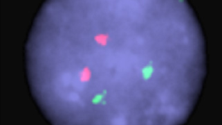
In an abnormal cell containing the t(11;14), one orange (CCND1/MYEOV), one green (IGH), and two fusion signal pattern (der (11) and der (14)) may be observed. Some samples containing the t(11;14) may display signal patterns different than one orange, one green, and two fusions.
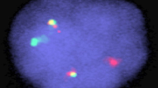
In a normal cell that lacks the t(11;14), a two orange and two green signal pattern will be observed reflecting the two intact copies of CCND1/MYEOV and IGH respectively.
Normal Hybridization: A normal interphase cell hybridized with the Vysis LSI IGH/CCND1 XT Dual Color Dual Fusion Probes. The cell shows the expected two orange (CCND1/MYEOV), two green (IGH) signal pattern.
Abnormal Hybridization: An abnormal interphase cell hybridized with the Vysis LSI IGH/CCND1 XT Dual Color Dual Fusion Probes. The cell in this image shows the one orange (CCND1/MYEOV), one green (IGH), two fusion (der (11) and der (14)) signal pattern indicative of a t(11;14).

Please be aware that the website you have requested is intended for the residents of a particular country or countries, as noted on that site. As a result, the site may contain information on pharmaceuticals, medical devices and other products or uses of those products that are not approved in other countries or regions.
Please be aware that the website you have requested is intended for the residents of a particular country or countries, as noted on that site. As a result, the site may contain information on pharmaceuticals, medical devices and other products or uses of those products that are not approved in other countries or regions.