MULTIPLE MYELOMA
Vysis LSI IGH/FGFR3 DF FISH Probe Kit
For more information, contact Abbott.
For more information, contact Abbott.
| PRODUCT NAME | UNIT SIZE | ORDER NUMBER | GTIN |
|---|
The Vysis IGH/FGFR3 DF FISH Probe Kit is intended to detect the t(4;14) (p16;q32) reciprocal translocation involving the FGFR3 and IGH gene regions.
The t(4;14)(p16;q32) is a common translocation in multiple myeloma (MM), but often missed by cytogenetics due to the telomeric chromosomal location of the regions involved in the translocation. The IFM99 trial demonstrates that the t(4;14) negatively impacted both event-free survival and overall survival in newly diagnosed symptomatic myeloma patients. FISH testing for t(4;14)( p16;q32) has been indicated as one of the minimum clinical tests during MM diagnosis and treatment determination.
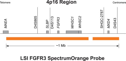
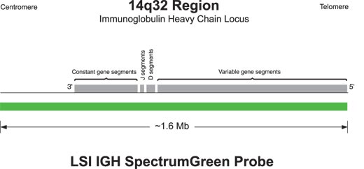
The SpectrumOrange probe is composed of 2 segments that span approximately 1Mb (UCSC March 2006 location is chr4:1300606- 2321488) with an approximately 39 kb gap (UCSC March 2006 location is chr4:2043467-2082355). The approximately 1.6 Mb SpectrumGreen probe spans the IGH region. (UCSC March 2006 location is chr14:104736507-106339460).
In a normal cell that lacks the t(4;14), a two orange and two green signal pattern will be observed reflecting the two intact copies of the FGFR3 and IGH regions respectively. In an abnormal cell containing the t(4;14), one orange (FGFR3), one green (IGH), and two fusion signalpattern (der (4) and der (14)) may be observed. Some samples containing the t(4;14) may display signal patterns differently than one orange, one green, and two fusions.
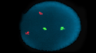
Normal Hybridization: An interphase cell hybridized with the LSI IGH/FGFR3 Dual Color Dual Fusion Probe. The cell shows the two orange (FGFR3),two green (IGH) signal pattern.
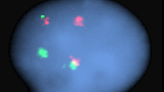
Abnormal Hybridization: An interphase cell hybridized with the LSI IGH/FGFR3 Dual Color, Dual Fusion Probe. The cell in this image shows the one orange (FGFR3), one green (IGH), two fusion (der (4) and der (14)) signal pattern.

Please be aware that the website you have requested is intended for the residents of a particular country or countries, as noted on that site. As a result, the site may contain information on pharmaceuticals, medical devices and other products or uses of those products that are not approved in other countries or regions.
Please be aware that the website you have requested is intended for the residents of a particular country or countries, as noted on that site. As a result, the site may contain information on pharmaceuticals, medical devices and other products or uses of those products that are not approved in other countries or regions.