MULTIPLE MYELOMA
Vysis LSI IGH/MAF DF FISH Probe Kit
For more information, contact Abbott.
For more information, contact Abbott.
| PRODUCT NAME | UNIT SIZE | ORDER NUMBER | GTIN |
|---|
The Vysis IGH/MAF DF FISH Probe Kit is intended to detect the t(14;16)(q32;q23) reciprocal translocation involving the IGH and MAF gene regions.
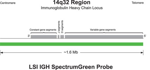
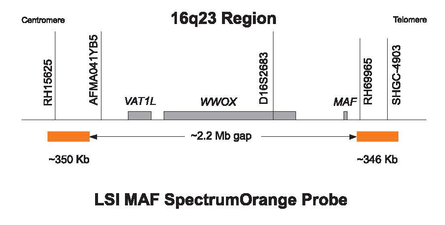
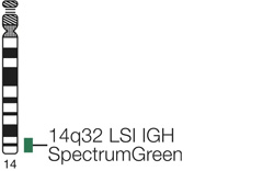
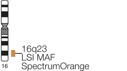
The SpectrumOrange probe flanks the MAF gene region and is composed of 2 segments that are each approximately 350 kb with an approximately 2.2 Mb gap. The centromeric segment is located at chr16:75729985-76079705 (March 2006 assembly, UCSC Genome Browser) and the telomeric segment is located at chr16:78290003-78635873 (March 2006 assembly, UCSC Genome Browser). The approximately 1.6 Mb SpectrumGreen probe spans the IGH region. (chr14:104736507-106339460; March 2006 assembly, UCSC Genome Browser)
In an abnormal cell containing the t(14;16), one green (IGH), one orange (MAF) and two fusion signal pattern (der (14) and der (16)) may be observed. Some samples containing the t(14;16) may display signal patterns different than one orange, one green and two fusions.
In a normal cell that lacks the t(14;16), a two green and two orange signal pattern will be observed reflecting the two intact copies of IGH and the MAF region respectively. Due to the presence of the ~2.2 Mb gap between the two SpectrumOrange labeled MAF probes, signal splitting of the orange probe may be observed in both normal and abnormal cells.
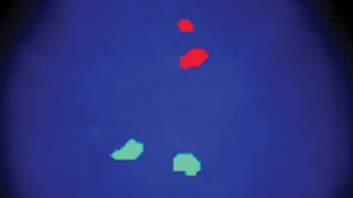
Normal Hybridization: An interphase cell hybridized with the LSI IGH/MAF Dual Color, Dual Fusion Translocation Probe. The cell shows the two green (IGH), two orange (MAF) signal pattern.
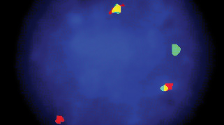
Abnormal Hybridization: An abnormal interphase cell hybridized with the Vysis LSI IGH/CCND1 XT Dual Color Dual Fusion Probes. The cell in this image shows the one orange (CCND1/MYEOV), one green (IGH), two fusion (der (11) and der (14)) signal pattern indicative of a t(11;14).

Please be aware that the website you have requested is intended for the residents of a particular country or countries, as noted on that site. As a result, the site may contain information on pharmaceuticals, medical devices and other products or uses of those products that are not approved in other countries or regions.
Please be aware that the website you have requested is intended for the residents of a particular country or countries, as noted on that site. As a result, the site may contain information on pharmaceuticals, medical devices and other products or uses of those products that are not approved in other countries or regions.