For more information, contact Abbott.
For more information, contact Abbott.
| PRODUCT NAME | UNIT SIZE | ORDER NUMBER | GTIN |
|---|
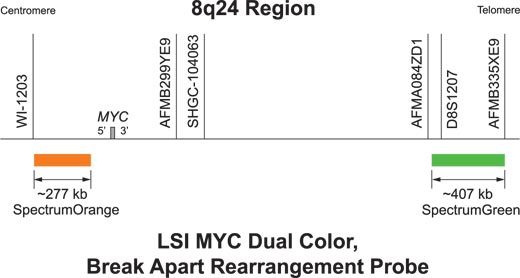
The Vysis LSI MYC Dual Color Break Apart Rearrangement fluorescence in situ hybridization probe is intended to detect chromosomal rearrangements involving the MYC gene region on chromosome 8q24. It is particularly useful for detection of aberrations with breakpoints located far telomeric to MYC such as those that can occur in the variant t(8;22)(q24.1;q11.2) IGL-MYC and t(2;8)(p11.2;q24.1) IGK-MYC rearrangements.
Translocations involving the MYC region have diagnostic and prognostic importance in B-cell malignancies. In Burkitt’s lymphoma approximately 75 to 80% of cases carry t(8;14) IGH-MYC and the remainder are associated with t(8;22) IGL-MYC or t(2;8) IGK-MYC. In approximately 5 to 10% of diffuse large B-cell lymphoma (DLBCL) patients also have MYC region rearrangements, and detection of these rearrangements with the MYC Dual Color Break Apart Rearrangement Probe has been associated with a poor prognosis. It has been suggested that FISH analysis for MYC rearrangements should be performed on all DLBCL patients.
The approximately 277 kb (chr8:128432540-128709819; March 2006 assembly)4 SpectrumOrange probe starts 119 kb centromeric to the MYC gene and is centromeric to the common breakpoint region. The approximately 407 kb (chr8:130338931-130745615; March 2006 assembly)4 SpectrumGreen probe begins approximately 1.5 Mb telomeric to the MYC gene and is telomeric to the breakpoint region observed in t(8;22) and t(2;8) translocations.
An abnormal nucleus hybridized with the LSI MYC Dual Color Break Apart Rearrangement Probe produces a two orange/green (yellow) fusion (2F) pattern. A one orange, one green, and one fusion pattern (1O1G1F) is expected from a sample with a t(2;8), t(8;22) or t(8;14) having a breakpoint within the gap between the hybridization targets of the LSI MYC probes
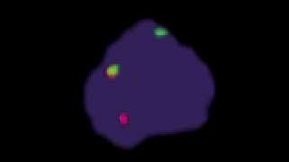
Abnormal Hybridization: LSI MYC Dual Color Break Apart Rearrangement Probe hybridized to an abnormal nucleus showing a one orange, one green and one orange/ green fusion (1O1G1F) signal pattern.

Please be aware that the website you have requested is intended for the residents of a particular country or countries, as noted on that site. As a result, the site may contain information on pharmaceuticals, medical devices and other products or uses of those products that are not approved in other countries or regions.
Please be aware that the website you have requested is intended for the residents of a particular country or countries, as noted on that site. As a result, the site may contain information on pharmaceuticals, medical devices and other products or uses of those products that are not approved in other countries or regions.