ACUTE MYELOGENOUS LEUKEMIA (AML)
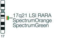
Vysis LSI RARA Dual Color Break Apart Rearrangement Probe
For more information, contact Abbott.
For more information, contact Abbott.
| PRODUCT NAME | UNIT SIZE | ORDER NUMBER | GTIN |
|---|
The Vysis RARA Break Apart FISH Probe is intended to detect chromosomal rearrangements involving the RARA gene region at chromosome 17q21 using the fluorescence in situ hybridization (FISH) technique.
Acute promyelocytic leukemia (APL) is associated with chromosomal rearrangements involving the retinoic acid receptor μ (RARA) gene on chromosome 17q21 and variable partner genes. In the vast majorityof APL cases, the RARA gene fuses with the promyelocytic leukemia gene (PML) located on chromosome 15q22 resulting in a t(15;17) translocation. RARA fusions with promyelocytic leukemia zinc finger (PLZF, 11q13), nucleophosmin (NPM, 5q35), nuclear mitotic apparatus (NuMA,11q23), signal transducer and activator of transcription 5b (STAT5B, 17q21), and PRKAR1A (protein kinase, cAMP- dependent, regulatory, type I, alpha, 17q23-q24) genes are also described.
The Vysis RARA Break Apart FISH Probe Kit has been used in several studies to detect chromosome 17q21 rearrangements involving the RARA gene.
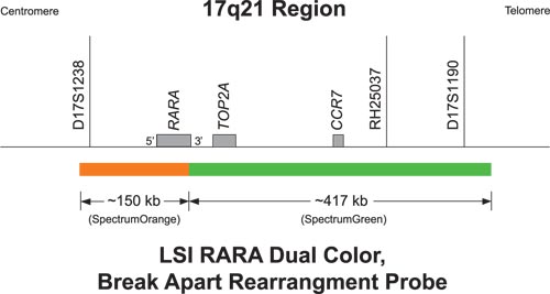
The approximately 150 kb (chr17:35612650-35762683; March 2006 assembly, UCSC Genome Browser) SpectrumOrange probe lies mostly centromeric to the RARA gene breakpoint region which occurs in intron 2. The probe does extend about 4 kb telomeric beyond intron 2. The approximately 417 kb (chr17:35762877-36180271; March 2006 assembly, UCSC Genome Browser) SpectrumGreen probe lies telomeric to the RARA breakpoint region.
The signal pattern observed in a cell that is lacking a RARA gene rearrangement consists of two orange/green (yellow) fusion signals (2F). The two fusion signals represent the normal (non-rearranged) RARA genes located on both 17 chromosomes. A signal pattern indicative of the RARA gene rearrangement is one orange, one green, and one green/orange (yellow) fusion signal. The separation of orange and green signals from one fusion (1O1G1F) indicates that the RARA gene has split apart.
The remaining single fusion signal represents the normal (non- rearranged RARA) gene on the normal chromosome extends approximately 400 kb toward the telomere of chromosome 17.
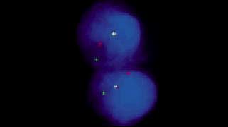
Abnormal Hybridization: Vysis LSI RARA Dual Color, Break Apart Rearrangement Probe hybridized to nuclei containing one orange, one green and one fusion (1O1G1F) signal pattern.

Please be aware that the website you have requested is intended for the residents of a particular country or countries, as noted on that site. As a result, the site may contain information on pharmaceuticals, medical devices and other products or uses of those products that are not approved in other countries or regions.
Please be aware that the website you have requested is intended for the residents of a particular country or countries, as noted on that site. As a result, the site may contain information on pharmaceuticals, medical devices and other products or uses of those products that are not approved in other countries or regions.