For more information, contact Abbott.
For more information, contact Abbott.
| PRODUCT NAME | UNIT SIZE | ORDER NUMBER | GTIN |
|---|
These fluorescence in situ hybridization (FISH) probes are intended to detect the t(8;21)(q21.3;q22) reciprocal translocation involving the RUNX1 and RUNX1T1 gene regions.
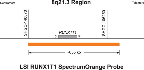
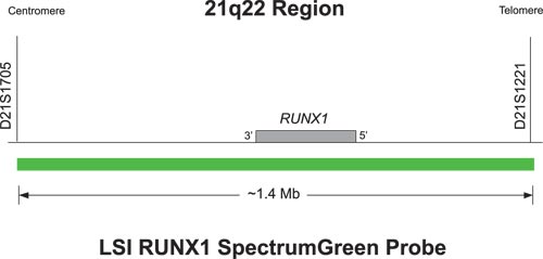
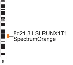
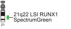
The approximately 1.4 Mb SpectrumGreen probe spans the RUNX1 gene (chr21:34452353-35813329; March 2006 assembly, UCSC Genome Browser). The approximately 655 kb SpectrumOrange probe spans the RUNX1T1 gene (chr8:92827265-93482325; March 2006 assembly, UCSC Genome Browser).
In a normal cell without the RUNX1/RUNX1T1 (also called AML1/ETO) fusion gene, two orange signals representing normal copies of RUNX1T1 and two green signals representing normal copies of RUNX1 are observed. In a cell containing the RUNX1/RUNX1T1 fusion gene, one orange (RUNX1T1), one green (RUNX1), and two orange/green (yellow) fusion signals are observed. The fusion signals represent the juxtaposition of the translocated portions of the two gene regions on the der(8) and the der(21). Variant RUNX1/RUNX1T1 signal patterns other than the most commonly observed one orange, one green, and two fusions (1O1G2F), may also occur.

Abnormal Hybridization: Vysis LSI RUNX1/RUNX1T1 Dual Color Dual Fusion Probes hybridized to an abnormal nucleus showing a one orange, one green and two fusion (1O1G2F) signal pattern.

Please be aware that the website you have requested is intended for the residents of a particular country or countries, as noted on that site. As a result, the site may contain information on pharmaceuticals, medical devices and other products or uses of those products that are not approved in other countries or regions.
Please be aware that the website you have requested is intended for the residents of a particular country or countries, as noted on that site. As a result, the site may contain information on pharmaceuticals, medical devices and other products or uses of those products that are not approved in other countries or regions.