Vysis LSI IGH/MAF Dual Color Dual Fusion Probe
For more information, contact Abbott.
For more information, contact Abbott.
| PRODUCT NAME | UNIT SIZE | ORDER NUMBER | GTIN |
|---|
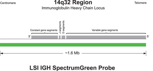
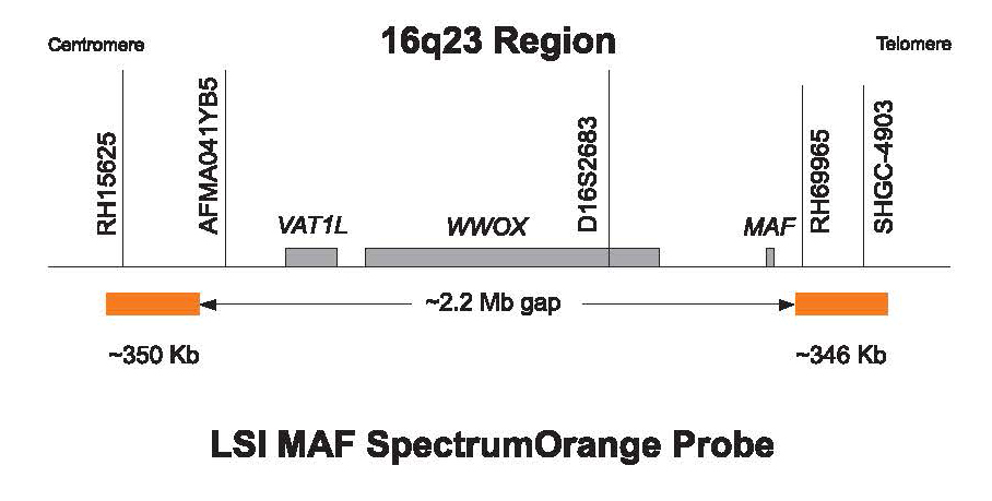
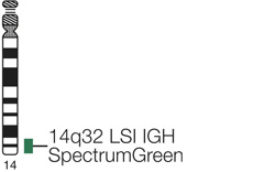
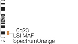
Vysis LSI IGH/MAF Dual Color Dual Fusion Probes hybridize to chromosome 14q32 (IGH SpectrumGreen) and chromosome 16q23 (MAF SpectrumOrange). The hybridized probe fluoresces with moderate to bright intensity both in interphase nuclei and on metaphase chromosomes.
In an abnormal cell containing the t(14;16), one green (IGH), one orange (MAF) and two fusion signal pattern (der (14) and der (16)) may be observed. Some samples containing the t(14;16) may display signal patterns different than one orange, one green and two fusions. In a normal cell that lacks the t(14;16), a two green and two orange signal pattern will be observed reflecting the two intact copies of IGH and the MAF region respectively. Due to the presence of the ~2.2 Mb gap between the two SpectrumOrange labeled MAF probes, signal splitting of the orange probe may be observed in both normal and abnormal cells.

Normal Hybridization: An interphase cell hybridized with the LSI IGH/MAF Dual Color, Dual Fusion Translocation Probe. The cell shows the two green (IGH), two orange (MAF) signal pattern.
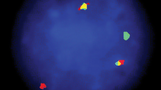
Abnormal Hybridization: An interphase cell hybridized with the LSI IGH/MAF Dual Color, Dual Fusion Translocation Probe. The cell in this image shows the one green (IGH), one orange (MAF), two fusion (der (14) and der (16)) signal pattern indicative of a y(14;16)

Please be aware that the website you have requested is intended for the residents of a particular country or countries, as noted on that site. As a result, the site may contain information on pharmaceuticals, medical devices and other products or uses of those products that are not approved in other countries or regions.
Please be aware that the website you have requested is intended for the residents of a particular country or countries, as noted on that site. As a result, the site may contain information on pharmaceuticals, medical devices and other products or uses of those products that are not approved in other countries or regions.